Surgery · Twelve procedures
Major
Surgeries
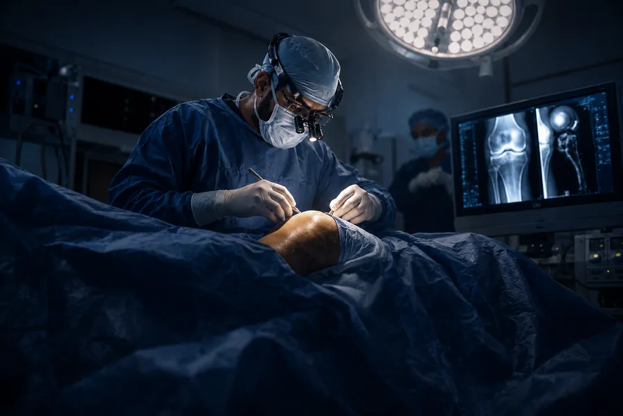
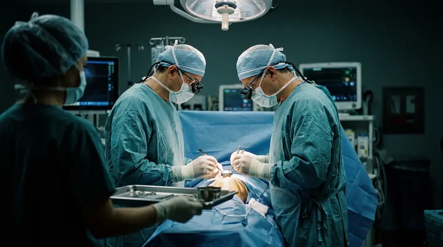
Joint replacement, complex trauma and reconstructive surgery, planned and performed in our own operation theatres at Vishwas Orthopaedic Hospital, Bharuch.
What this hospitalis built to treat.
Four areas carry most of our surgical work. Each is supported by our own operation theatres, imaging, implant inventory and physiotherapy unit.
01
Complex trauma & polytrauma care
Advanced surgical management of high-energy road traffic accidents, compound (open) fractures, pelvi-acetabular injuries and peri-articular fractures — the injuries where the first operation decides the final result.
02
Primary & revision joint replacement
Total and partial replacement of the knee and hip for severe degenerative arthritis, avascular necrosis (AVN) and secondary arthritis, including complex and revision arthroplasty when an earlier implant has failed.
03
Geriatric fracture management
Minimally invasive stabilisation tailored to osteoporotic bone, chosen so that older patients stand and walk early and avoid the complications that come from weeks in bed.
04
Deformity correction & reconstruction
Management of long-bone malunions that have healed crooked and non-unions that have not healed at all, alongside salvage reconstruction for bone loss and infection.
Twelve major surgeries,in four families.
Jump straight to the group you are looking for. Every procedure below is described in plain language, with what it treats and what recovery looks like.
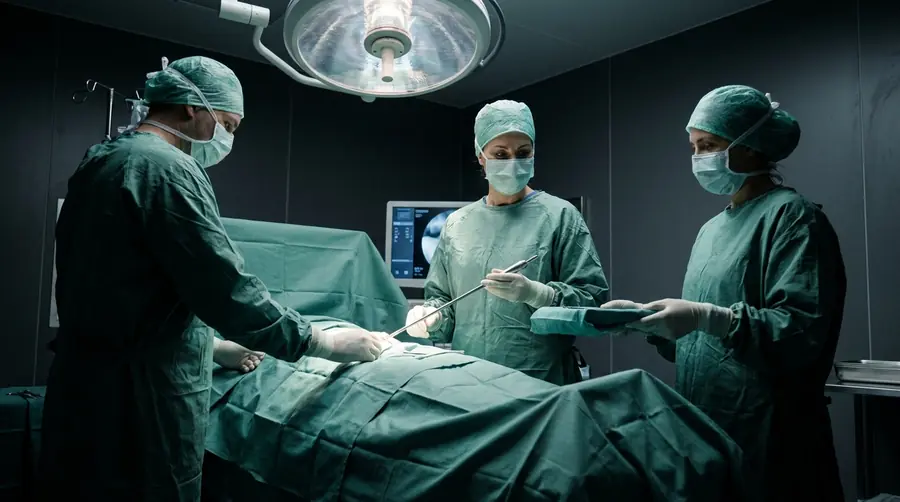
Joint Replacement
Four procedures — knee, hip, partial and revision replacement for arthritis, AVN and failed implants.
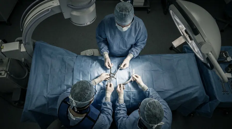
Complex Trauma
Three procedures — pelvic and acetabular fixation, joint-surface reconstruction and polytrauma stabilisation.
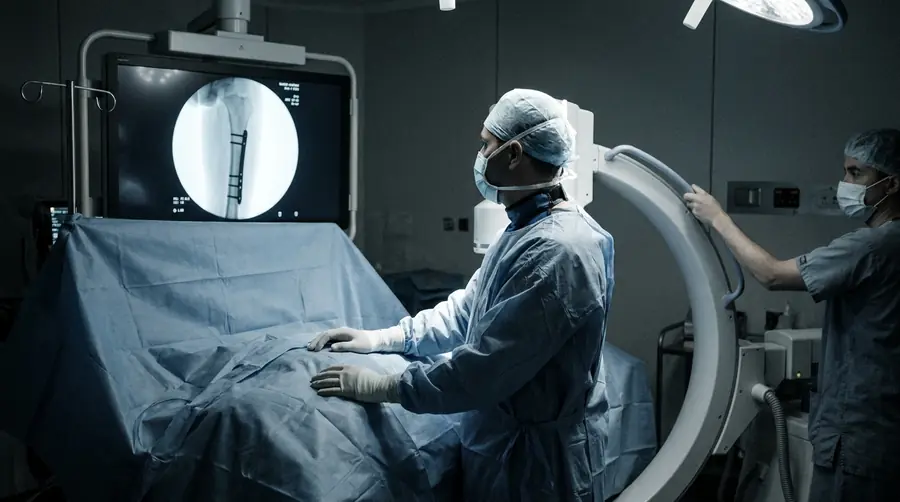
Minimally Invasive
Two procedures — percutaneous plating and closed interlocking nailing through small incisions.
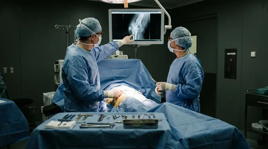
Salvage & Reconstruction
Three procedures — non-union and malunion correction, revision fixation and infection surgery.
Joint replacement(arthroplasty).
Replacement is offered when the joint surface itself is gone — not before. When it is the right answer, it is one of the most reliable operations in orthopaedics.
01
Knee
Total Knee Replacement (TKR)
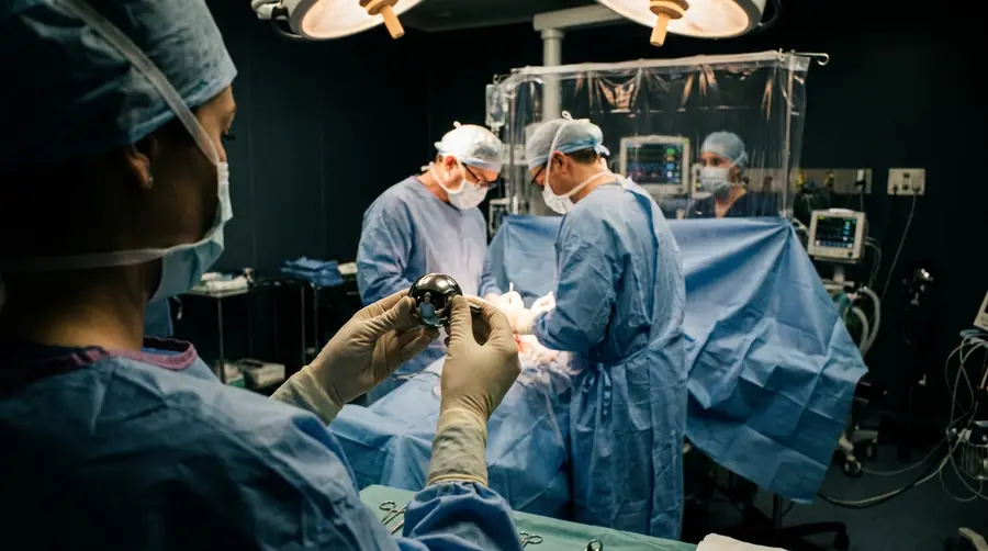
The worn surfaces of the knee are removed and replaced with a metal and polyethylene implant, aligned to your own limb axis rather than a standard template. Offered for advanced degenerative arthritis, post-traumatic and secondary arthritis, and deformity that no longer answers to medication, injections or physiotherapy. Standing and assisted walking usually begin on the first day after surgery.
02
Hip
Total Hip Replacement (THR)
Both the femoral head and the socket are replaced, restoring a smooth, stable and correctly tensioned hip joint. The common reasons are advanced hip arthritis, avascular necrosis (AVN) of the femoral head and arthritis that follows an old injury. Implant choice and approach are decided from your own radiographs and your age, bone quality and activity.
03
Hip · Fracture
Partial / Hemi-Arthroplasty (Bipolar Hip Replacement)
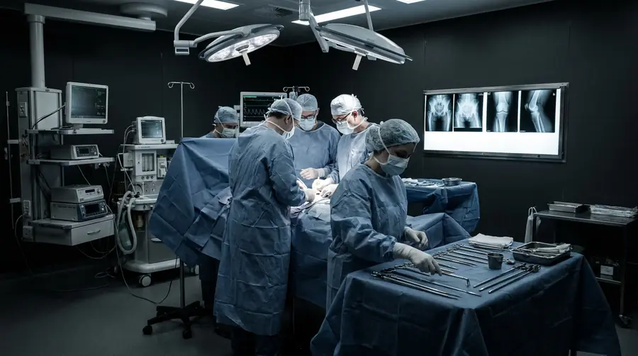
For a displaced fracture of the neck of the femur — most often in an older patient with osteoporotic bone — the fractured head is replaced with a bipolar prosthesis instead of being fixed and left to unite unreliably. The aim is deliberate and specific: get the patient upright and walking within a day or two, because prolonged bed rest is itself dangerous at that age.
04
Revision
Complex & Revision Knee and Hip Replacement
When an earlier replacement loosens, wears out, becomes unstable or becomes infected, the old implant is removed and the joint is rebuilt — often with longer stems, metal augments and bone grafting to replace what has been lost. These are longer, more demanding operations, planned in advance with imaging and a full implant inventory kept ready.
Complex traumasurgery.
High-energy injuries, open fractures and fractures that enter a joint. These are the operations the hospital was equipped for first.
05
Pelvis
Pelvic and Acetabular Fracture Fixation
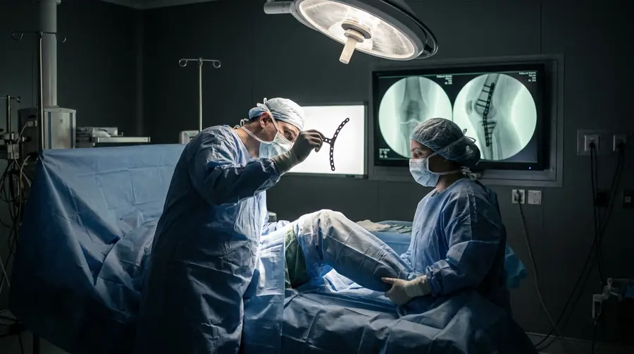
Fractures of the pelvic ring and the hip socket follow high-energy injury — road traffic accidents and falls from height. They are among the most technically demanding fractures in the body. Reduction and fixation with contoured reconstruction plates restore the weight-bearing dome of the socket and the stability of the ring, which is what protects the hip from early arthritis later.
06
Joint surface
Intra-Articular & Peri-Articular Fracture Reconstruction
Fractures that run into a joint — distal femur, tibial plateau, pilon of the ankle and proximal humerus — are reconstructed so that the joint surface is anatomically restored and held with anatomically contoured locking plates. Millimetres matter here: a step in the joint surface becomes stiffness and arthritis within a few years.
07
Polytrauma
Polytrauma Stabilisation & Damage Control Orthopaedics (DCO)
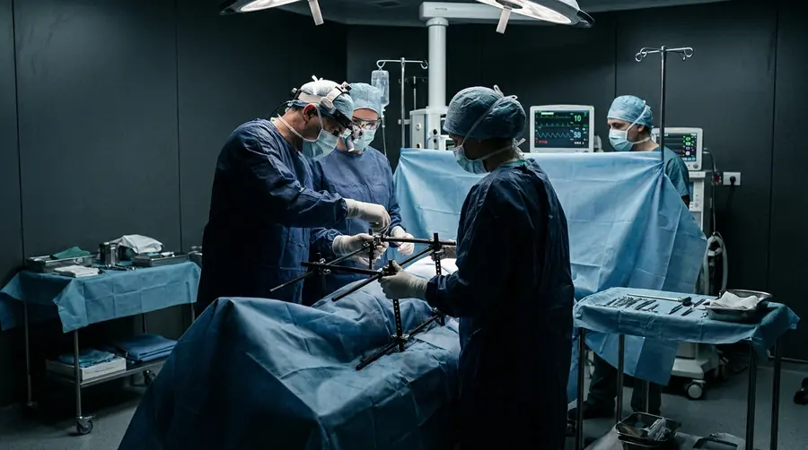
When a patient arrives with several major injuries at once, survival comes before definitive fixation. Bleeding is controlled, fractures are stabilised quickly with external fixators or traction, and the patient is allowed to recover physiologically. Definitive internal fixation follows in a planned second stage, once it is safe — that sequence is what damage control orthopaedics means.
Minimally invasivetrauma surgery.
Fixing the fracture without destroying the tissue around it. Smaller incisions, preserved blood supply, faster and more dependable union.
08
MIPPO
Minimally Invasive Percutaneous Plate Osteosynthesis
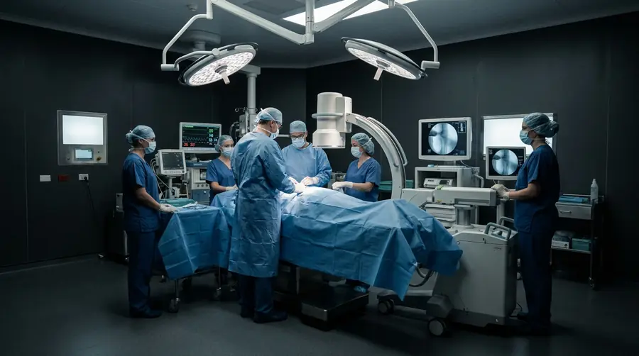
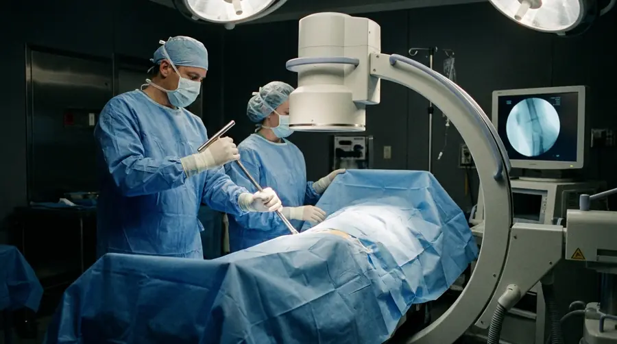
The plate is slid along the bone beneath the muscle through two small windows, and locked in place under fluoroscopic guidance, without opening and stripping the fracture itself. The fracture’s own blood supply and the surrounding soft tissue envelope are left largely undisturbed, which means less pain, less blood loss, a lower infection risk and more reliable healing.
09
Nailing
Closed Interlocking Intramedullary Nailing
For fractures of the shaft of the femur, tibia and humerus, a titanium nail is passed down the medullary canal through a small incision away from the fracture and locked with screws at both ends. The fracture is never exposed. Because the nail shares load with the bone, controlled weight bearing can often start early, and the scar is a fraction of the size of an open plating scar.
Salvage &reconstructive surgery.
The second-chance operations — for fractures that failed to heal, fixation that failed to hold, and bone that became infected.
10
Non-union
Non-Union and Malunion Corrective Surgery, with Bone Grafting
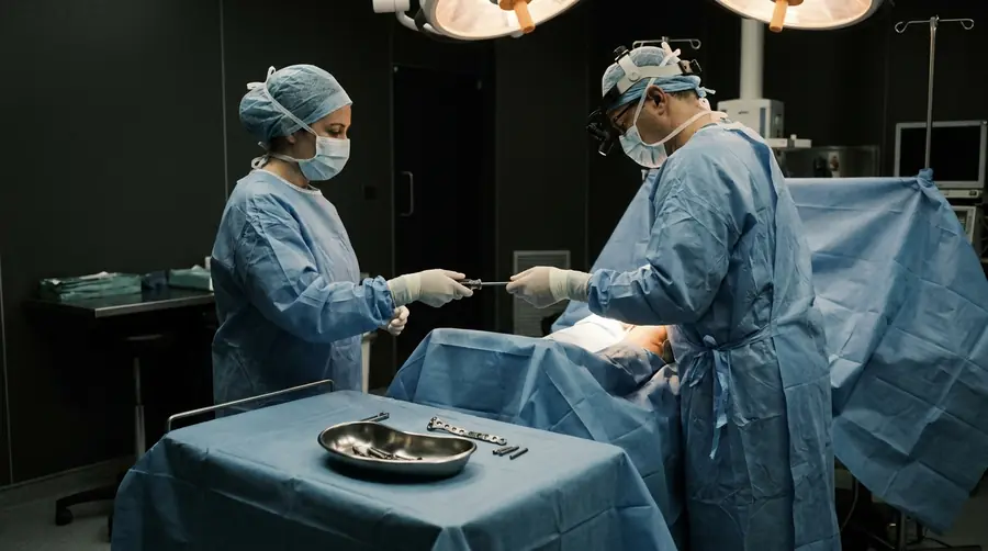
Some fractures never unite, and some unite in the wrong position. Both are correctable. The deformity is cut and realigned through a planned osteotomy, stable fixation is applied, and cancellous bone graft — usually taken from the patient’s own iliac crest — is packed at the site to restart biological healing.
11
Revision fixation
Implant Removal and Revision Fixation
Hardware is removed when it has done its job and become a source of irritation, or when it has failed — a broken plate, backed-out screws, a fixation that has lost hold in soft bone. Where the fracture is still unhealed, removal is combined with revision fixation using a different and more suitable construct.
12
Infection
Chronic Osteomyelitis Debridement & Reconstruction
Long-standing bone infection is treated surgically, not with antibiotics alone. Dead and infected bone is debrided back to bleeding tissue, local antibiotic delivery is used to sterilise the bed, and the resulting defect is reconstructed in a planned staged procedure once the infection is controlled.
Everything we treat,not only what we operate.
Most people who walk into the OPD never reach an operation theatre. These are the treatments and services available across the hospital.
01 — Emergency care
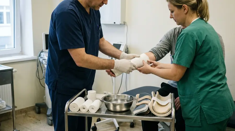
Emergency trauma & fracture care
- Round-the-clock management of simple, comminuted and open fractures
- Closed reduction and cast or slab immobilisation
- Soft tissue injury management, wound care and dressings
- Dislocation reduction and splinting
02 — Fixation
Advanced internal & external fixation
- Minimally invasive plate osteosynthesis (MIPO / MIPPO)
- Interlocking intramedullary nailing
- Tension band wiring
- External fixator application for limb-threatening injuries
03 — Arthritis clinic
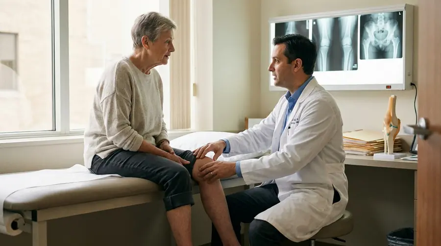
Joint care & arthritis clinic
- Medical management of osteoarthritis
- Intra-articular injections — viscosupplementation, corticosteroid and PRP therapy
- Tailored joint preservation protocols
- Assessment of when replacement is, and is not, the right answer
04 — Reconstruction
Post-traumatic reconstruction
- Surgery for non-healing fractures
- Bone grafting
- Infection eradication in chronic osteomyelitis
- Corrective osteotomies for malunion and deformity
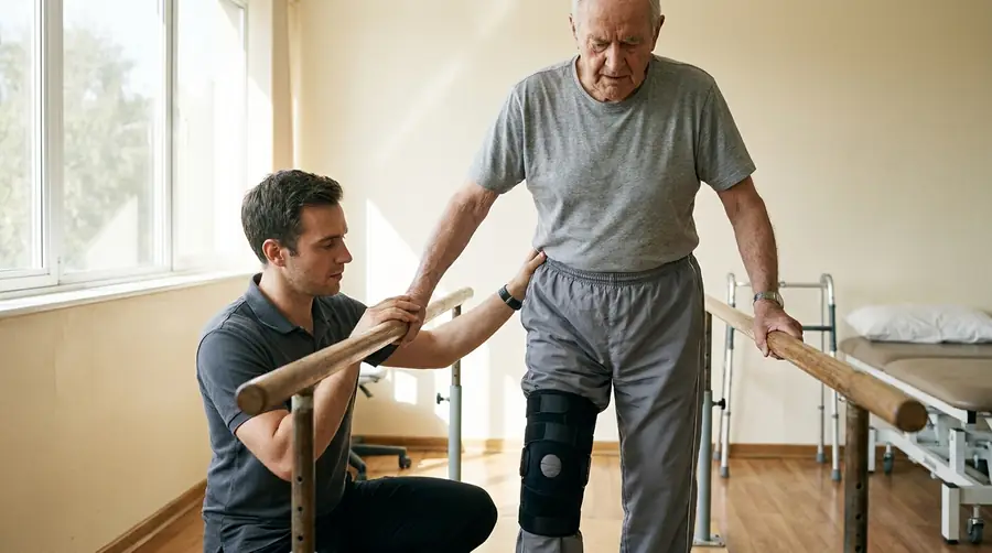
05 — Rehabilitation
Post-operative rehabilitation
- Structured, goal-oriented physiotherapy plans
- Restoring joint range of motion
- Muscle strengthening and gait retraining
- Progression to independent mobility
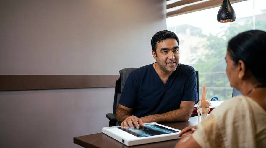
Bring the X-ray.We will tell you straight.
If surgery is not needed, we will say so. If it is, you will be told what it involves, what it costs you in recovery time, and what the alternatives are.
- OPD timings
- Monday to Saturday
Morning 10:30 – 13:30
Evening 17:30 – 19:30 - Phone
- +91 76004 62706
- +91 76004 62706
- @vishwas_ortho
- Emergency
- Trauma and fractures are attended round the clock. Where possible, call before arriving.
Before your consultation
- Carry every X-ray, CT or MRI you already have, including the old ones
- Bring your current medicine list and any implant or discharge papers
- Note down how far you can walk, and what movement hurts
- For a fracture, come as you are — do not wait for an appointment
Book a consultation WhatsApp us
Related reading: How long do fractures take to heal? →